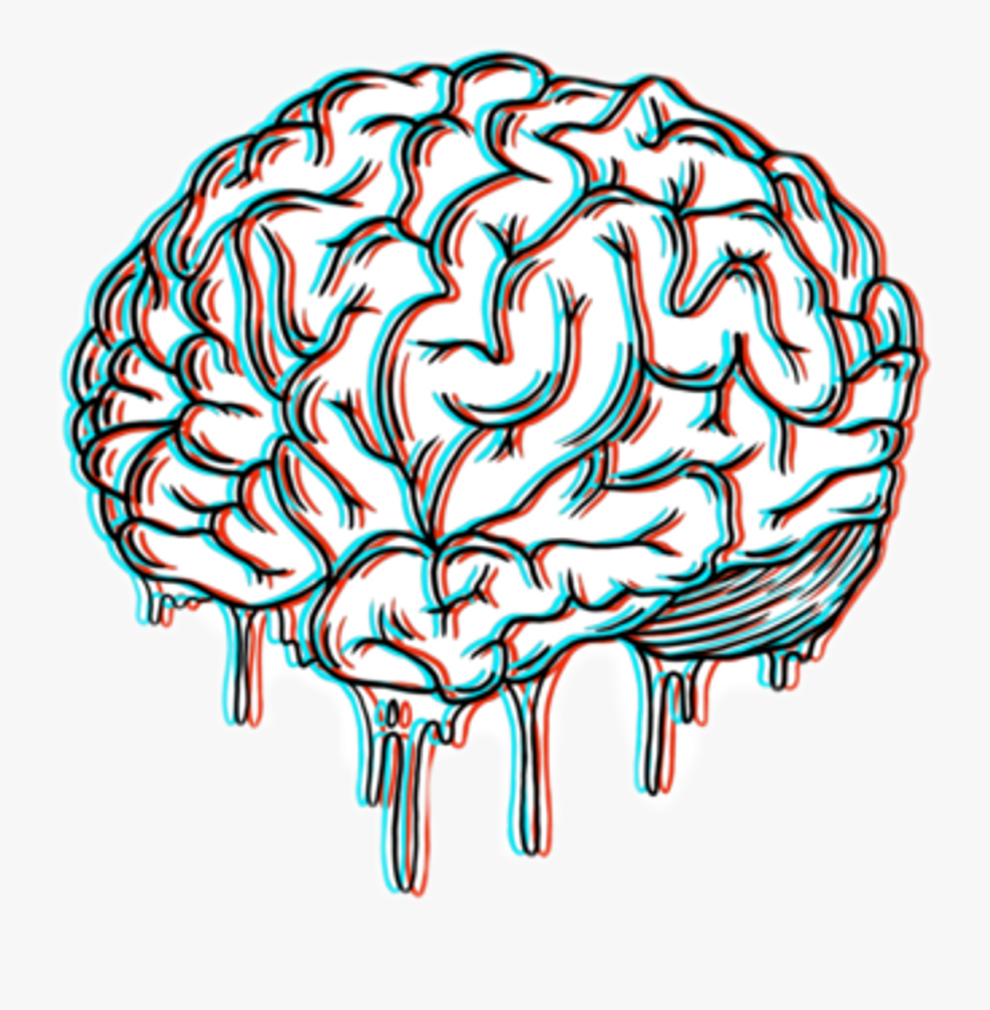
41 drawing of the brain with labels
Brain Hemisphere Hat – Ellen McHenry's Basement Workshop (The Science Museum in London used the brain hat as an adult activity. It’s also been used by a brain imaging company and several neuroscientists.) If you are using it with kids younger than 10 they will probably need help assembling it. Description of activity: Cut and assemble a paper brain hat. Time needed: ALL PROGRAMS MUST END The Senior Bulletin - Livonia Drawing the Audience into Your Performance Pt. 1 & 2, Scene Work-Study Pt 1. & 2, Auditions and Rehearsals, Pulling It All Together. ICE CREAM SOCIAL Wednesday, June 15, noon to 1 p.m. Our first ice cream social event is sponsored by our friends at American House of Livonia. They will prepare your sundae with all your
Image result for labeled diagram of the brain | Brain lobes, Human ... Mar 7, 2018 - Image result for labeled diagram of the brain. Mar 7, 2018 - Image result for labeled diagram of the brain. Pinterest. Today. Explore. ... There are uncharted worlds inside your head, but science is drawing a map. PennyDellPuzzles. Brain Health. Similar ideas popular now. Applied Science.

Drawing of the brain with labels
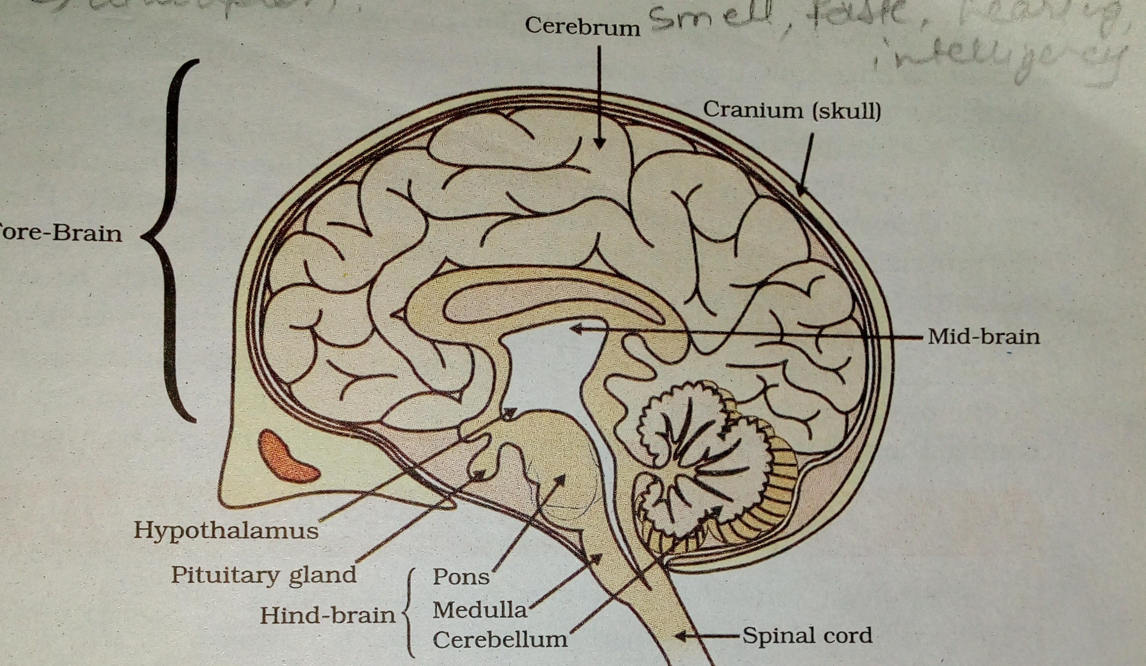
The brain and adrenal glands with hypothalamus, pituitary gland, and ... Drawing of the brain and adrenal glands with hypothalamus, pituitary gland, and adrenal glands labeled and arrows diagramming the effect of CRH on ACTH and the effect of ACTH on cortisol. Caption The hypothalamus sends CRH to the pituitary, which responds by secreting ACTH. ACTH then causes the adrenals to release cortisol into the bloodstream. The Allen Mouse Brain Common Coordinate Framework: A 3D ... May 14, 2020 · (N–R) Coronal STPT images of tdTomato reporter expression from the Slc17a8-IRES2-Cre line, which labels the ependymal cells lining the ventricles and central canal. Integration of these data types enabled the 3D reconstruction of ventricles and associated structures directly on the template volume. For all abbreviations see Table S2. Scale ... Solved 2. Draw a smple line diagram of the circle of Willis - Chegg 2. Draw a smple line diagram of the circle of Willis at the base of the brain by doing the following: a. Draw a line to show the division between the two hemispheres and label the top "frontal and the bottom occipital." b In the lower half of the drawing, show the basilar artery dividing into two posterior cerebral arteries, and extend each ...
Drawing of the brain with labels. Labeled Brain Model Diagram | Science Trends The cerebrum is the largest and most complex portion of the human brain. The cerebrum's function is to control our actions and thoughts, either conscious or unconscious, and responses to stimuli. The cerebrum itself is typically divided into four different lobes: the temporal lobe, the parietal lobe, the occipital lobe, and the frontal lobe. Labeled brain drawing Images, Stock Photos & Vectors - Shutterstock 4,937 labeled brain drawing stock photos, vectors, and illustrations are available royalty-free. See labeled brain drawing stock video clips Image type Orientation Color People Artists More Sort by Healthcare and Medical Clothing and Accessories Art Styles human brain anatomy t-shirt brain medicine organ logo Turn on AI Powered Search How to draw a The human Brain - YouTube How to draw a The human Brain easy and step by step. Draw this The human Brain by following this drawing lesson. Get The Markers HERE = What is Cognitive Neuroscience? Definition & FAQs | EMOTIV Cognitive Neuroscience Example. Examining cognitive neuroscience experiments is helpful to understand this subfield at work. A recent award-winning experiment explored the role of dopamine, a neurotransmitter associated with feelings of satisfaction, brain function, and decision making.
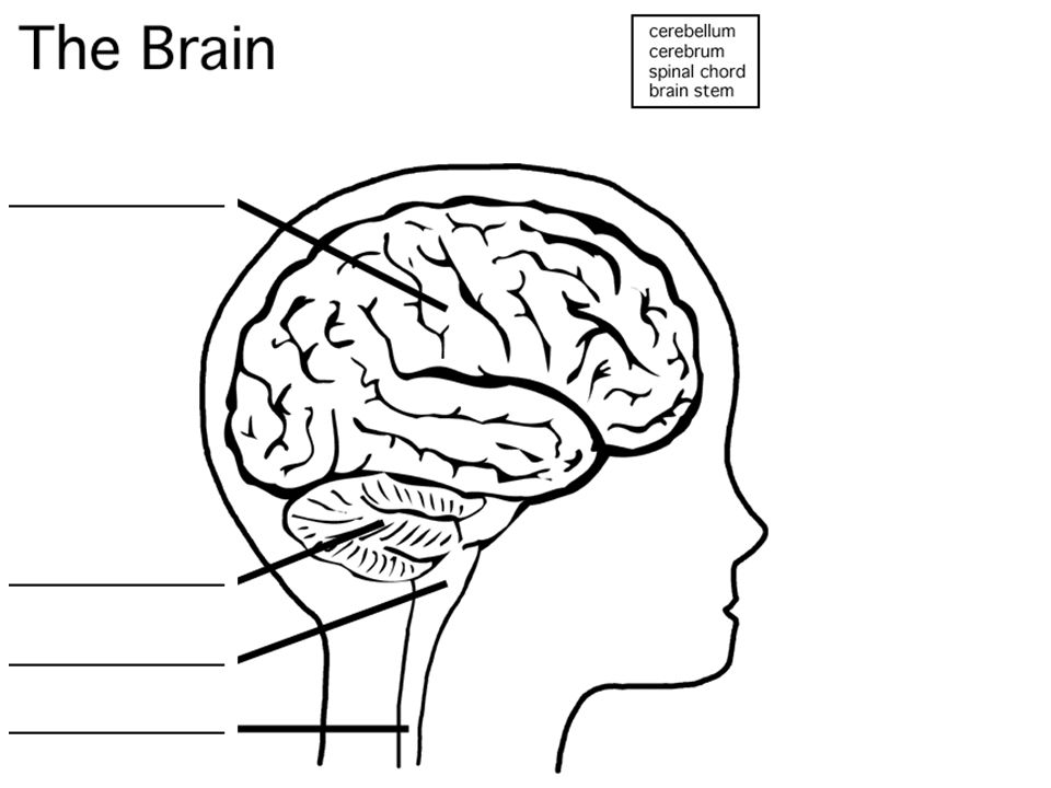
The Brain - Diagram and Explanation AMYGDALA: Lying deep in the center of the limbic emotional brain, this powerful structure, the size and shape of an almond, is constantly alert to the needs of basic survival including sex, emotional reactions such as anger and fear. Consequently it inspires aversive cues, such as sweaty palms, and has recently been associated with a range of mental conditions including depression to even autism. Anatomical diagrams of the brain - e-Anatomy - IMAIOS This anatomy module is about the anatomy of the central nervous system, especially the brain. It is composed of 64 drawings, illustrations and anatomical charts, all in "vector" format. ... use of interactive anatomical labels. The user can select to display multiple categories of labels on the illustrations: Cerebral lobes / regions; Cerebrum ... How to Draw a Brain: 14 Steps (with Pictures) - wikiHow Once you've drawn the cartoon or realistic brain, you can go back and add color or label the parts. Method 1 Sketching an Easy Cartoon Brain 1 Draw a large bean shape to make the outline for the brain. Use a pencil to sketch a kidney bean shape on your paper. You can make the outline for the brain any size you like. Answered: 1) Make a drawing of the nervous system a... |24HA 1) Make a drawing of the nervous system and label the brain, spinal cord, motor nerve and sensory nerve. 2) Provide an example of how the central nervous system and the peripheral nervous system work together to maintain homeostasis. As we exercise we create heat, in order to maintain a relatively constant core temperature the nervous system ...
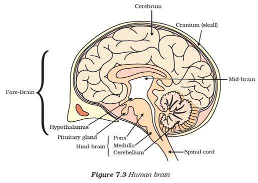
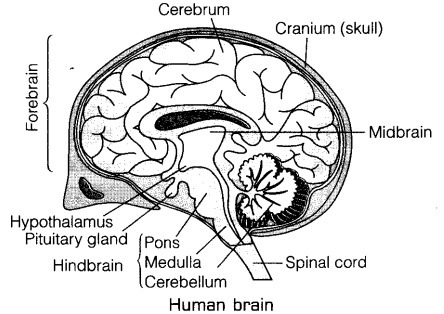
how to draw a brain - Google Search | Brain drawing, Brain art, Drawings This is a stunning watercolor print of an anatomically accurate human brain. The painting depicts the brain in a sagittal view, and shows parts of the temporal, parietal, frontal and occipital lobes, as well as the cerebellum. ---------------------- GO BIG! WITH LYON ROAD ART: Choose our larger size options to make an impact in your office or home. Left Brain vs. Right Brain: Characteristics Chart [INFOGRAPHIC] Oct 27, 2021 · Brain dominance theory is absorbing and enjoyable, plus it allows people to think about stereotypes and labels. In reality, though, psychology is complicated, and the truth is that there are very few people who have the traits of only one of these descriptions. Labeled Diagrams of the Human Brain You'll Want to Copy Now Labeled Diagrams of the Human Brain Central Core The central core consists of the thalamus, pons, cerebellum, reticular formation and medulla. These five regions are the central areas that regulate breathing, pulse, arousal, balance, sleep and early stages of processing sensory information. Diagram Of Brain with their Labelings and Detailed Explanation A well-labelled diagram of a human brain is given below for further reference. Structure And Function Of The Human Brain Parts Of The Human Brain The human brain is divided into three main parts: Forebrain. Midbrain. Hindbrain. These three main parts comprises many small parts. Forebrain The forebrain is also called as Prosencephalon.
Brain Drawing With Labels at GetDrawings | Free download
Brain Anatomy Labeled Stock Illustrations - Dreamstime Download 164 Brain Anatomy Labeled Stock Illustrations, Vectors & Clipart for FREE or amazingly low rates! New users enjoy 60% OFF. 188,693,165 stock photos online. ... Drawing of the skull showing the internal surface, with the posterior, middle and anterior fossae labeled.

Da Vinci's Wings: 4th Grade Self Portraits
How to draw and label human brain step by step.(full tutorial) hello friends in this video I have shown you how to draw human mind and label it step by step. friends I have explain all the parts of the human brain in thi...

Interesting Facts & Illustrations About the Brain Anatomy – Medical Stock Images Company
Happy Glass - Play Happy Glass on CrazyGames Happy Glass is a fun puzzle game featuring an empty glass that needs filling. Figure out how to get the water into the glass by drawing lines and make the glass happy again. The less you draw the higher your score.

Pablo Picasso Vallauris 1949 | modern design by moderndesign.org
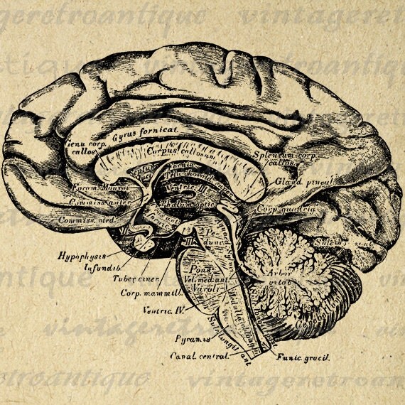
Solved Drawing of Mid-Sagittal Section of the Brain Label - Chegg Question: Drawing of Mid-Sagittal Section of the Brain Label the following: Midbrain Pons Medulla Cerebellum White Matter Gray Matter Thalamus Hypothalamus Third Ventricle Forth This problem has been solved!
Printable Image Brain Cross Section Digital by VintageRetroAntique
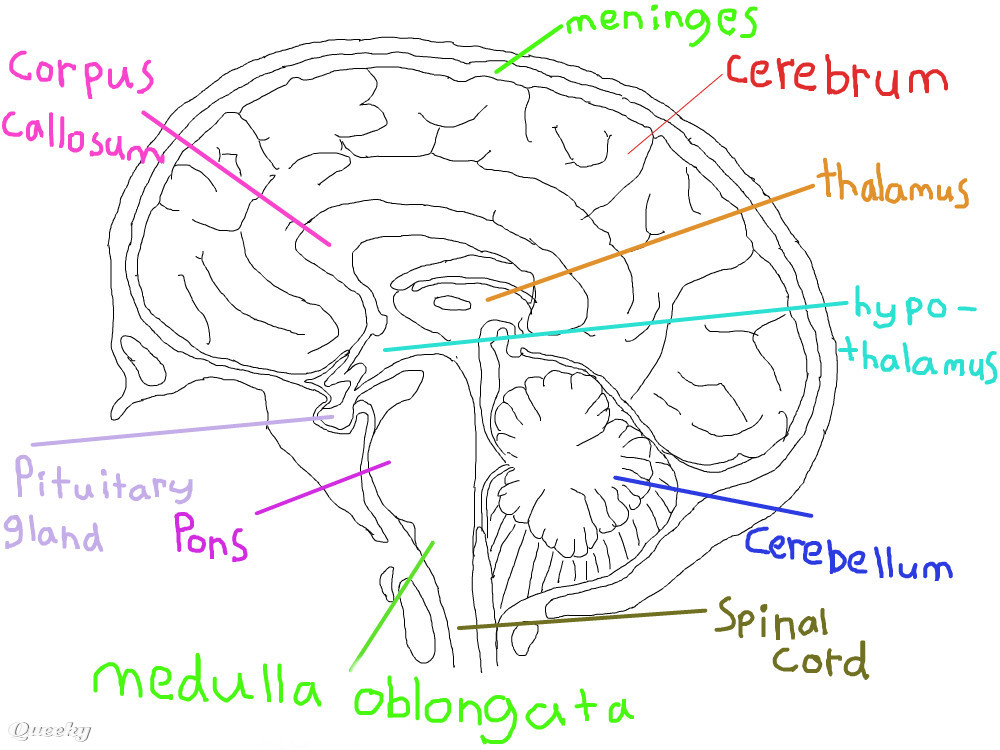
Label Brain Diagram Printout - EnchantedLearning.com The Brain. Read the definitions below, then label the brain anatomy diagram. Cerebellum - the part of the brain below the back of the cerebrum. It regulates balance, posture, movement, and muscle coordination. Corpus Callosum - a large bundle of nerve fibers that connect the left and right cerebral hemispheres.

Lab 2 Pig Heart-Labeled | Cardiovascular, Estudos
Diagram of the Brain and its Functions - Bodytomy Given below is a labeled diagram showing the brain stem and its related structures. Brain Stem and Structures Cerebellum The word 'cerebellum' literally means little brain. It is the second largest part of the brain, and is located at the back, below the occipital lobe, beneath the cerebrum and behind the brain stem.

Brain Viewed from Above | ClipArt ETC
Glucose and The Brain: Improving Mental Performance - Eufic Apr 30, 2013 · It does this through two main mechanisms: first, by drawing glucose directly from the blood when its cells are low on energy; and second, by limiting the amount of glucose available to the rest of the body so that there is more available to the brain. 2,3 These mechanisms are essential for survival. Unlike muscles (including the heart), and the ...

What is dyslexia? - Kelli Sandman-Hurley - YouTube
How to Draw A Brain : Step By Step Guide Step 1 Draw a simple oval for the cerebellum. Step 2 Now draw two geometrical shapes as shown for the two sides of the brain. Keep in mind narrower at the top. Step 3 Using the 2 shapes, draw the two sides of the brain as show with little pointed shape at the top. Step 4 Fill the brain with short and curved lines which shows millions of synapses.
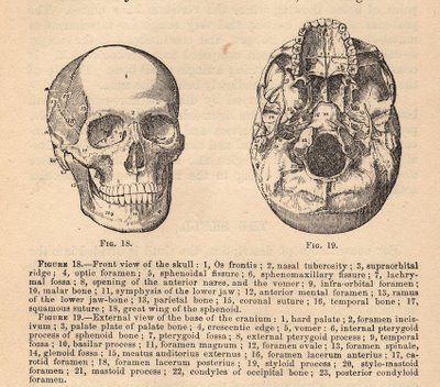
Vintage Graphic - Anatomy - Skull Diagram - The Graphics Fairy
Parts of the brain: Learn with diagrams and quizzes - Kenhub Labeled brain diagram. First up, have a look at the labeled brain structures on the image below. Try to memorize the name and location of each structure, then proceed to test yourself with the blank brain diagram provided below. Labeled diagram showing the main parts of the brain.
Brain Drawing With Labels at PaintingValley.com | Explore collection of Brain Drawing With Labels
101 Labeled Brain Images and a Consistent Human Cortical Labeling ... We selected 101 T 1-weighted brain MR images that are: (1) publicly accessible with a non-restrictive license, (2) from healthy participants, (3) of high quality to ensure good surface reconstruction, and (4) part of a multi-modal acquisition ( T 2*-weighted, diffusion-weighted scans, etc.).
Brain Clipart Simple Drawn - Aesthetic Brain Drawing , Free Transparent Clipart - ClipartKey
How to Draw a Human Brain - How to Draw Step by Step Drawing Tutorials (Step 1) Lightly draw a sideways "D"-like shape as a guide lines for drawing the brain. This shape will be erased later. (Step 2) Lightly draw an oval guide line. (Step 3) Lightly draw another oval guide line. Now we can start the real lines. (Step 4) Draw around the guide lines…make the line a little wavy, or not perfect, I mean.
Brain Drawing With Labels at GetDrawings | Free download
Draw Your Nervous System | AMNH Cut a sheet of paper that is longer than the height of your friend. (Or, tape sheets of paper together.) Place the paper on a hard, smooth floor. Have your friend lay down in the middle of the paper. Use a black marker to draw an outline of your friend's body. Next, draw the different parts of the nervous system. Then, label them.
Brain Drawing With Labels at PaintingValley.com | Explore collection of Brain Drawing With Labels
Draw labelled diagrams of the following: - Noon Academy Draw labelled diagrams of the following: (a) Neuron (b) Brain (c) Eye (d) Ear (a) Neuron (b) Brain (c) Eye (d)Ear. Search for: i. More Material. Write chromyl chloride test with equation. What do you understand by lanthanide contraction. What are lanthanide elements?

Pakmasti: Khana_Kaba_Masjid_e_nabvi
Nervous System - Label the Brain - TheInspiredInstructor.com This brain part controls thinking. This brain part controls balance, movement, and coordination. This brain part controls involuntary actions such as breathing, heartbeats, and digestion. This part of the nervous system moves messages between the brain and the body. This part of the cerebrum interprets and sorts information from the senses.
Drawing Of The Brain With Labels at GetDrawings.com | Free for personal use Drawing Of The Brain ...
How to Draw a Brain Step By Step - For Kids & Beginners How to draw a brain step by step: Step 1: Draw a bean shape with 3 curves at the lower end. Step 2: Draw 5 compartments inside the shape, as depicted below. Step 3: In the first compartment draw irregular lines. Step 4: Again draw random lines in the second compartment. Step 5: Similarly draw those lines in the 3rd compartment.

Reality Filter #3-4: The Upper Brain - Milestones San Diego Mommy and Me Classes
Solved 2. Draw a smple line diagram of the circle of Willis - Chegg 2. Draw a smple line diagram of the circle of Willis at the base of the brain by doing the following: a. Draw a line to show the division between the two hemispheres and label the top "frontal and the bottom occipital." b In the lower half of the drawing, show the basilar artery dividing into two posterior cerebral arteries, and extend each ...
Brain ← a tutorials Speedpaint drawing by Shleya - Queeky - draw & paint
The Allen Mouse Brain Common Coordinate Framework: A 3D ... May 14, 2020 · (N–R) Coronal STPT images of tdTomato reporter expression from the Slc17a8-IRES2-Cre line, which labels the ependymal cells lining the ventricles and central canal. Integration of these data types enabled the 3D reconstruction of ventricles and associated structures directly on the template volume. For all abbreviations see Table S2. Scale ...
Post a Comment for "41 drawing of the brain with labels"