42 images of compound microscope with labels
Compound Microscope- Definition, Labeled Diagram, Principle, Parts, Uses In order to ascertain the total magnification when viewing an image with a compound light microscope, take the power of the objective lens which is at 4x, 10x or 40x and multiply it by the power of the eyepiece which is typically 10x. Therefore, a 10x eyepiece used with a 40X objective lens will produce a magnification of 400X. › cemf › whatisemWhat is Electron Microscopy? - UMASS Medical School Conventional scanning electron microscopy depends on the emission of secondary electrons from the surface of a specimen. Because of its great depth of focus, a scanning electron microscope is the EM analog of a stereo light microscope. It provides detailed images of the surfaces of cells and whole organisms that are not possible by TEM.
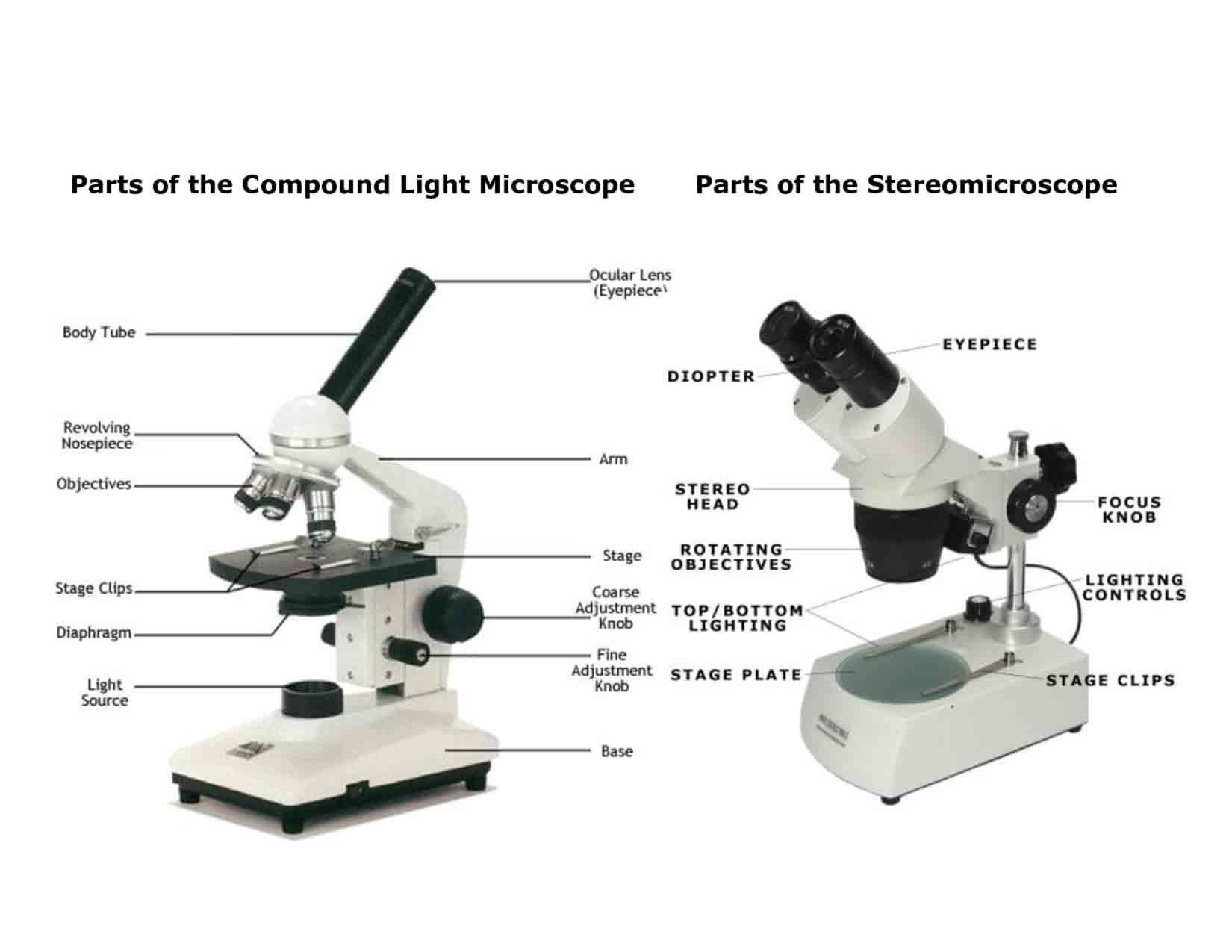
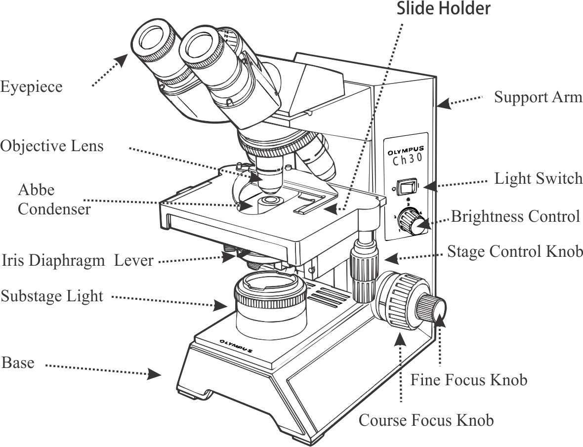
Compound Microscope Parts, Functions, and Labeled Diagram Compound Microscope Definitions for Labels. Eyepiece (ocular lens) with or without Pointer: The part that is looked through at the top of the compound microscope. Eyepieces typically have a magnification between 5x & 30x. Monocular or Binocular Head: Structural support that holds & connects the eyepieces to the objective lenses.

Images of compound microscope with labels
Microscope Parts and Functions First, the purpose of a microscope is to magnify a small object or to magnify the fine details of a larger object in order to examine minute specimens that cannot be seen by the naked eye. Here are the important compound microscope parts... Eyepiece: The lens the viewer looks through to see the specimen. What is a Compound Microscope? - Microscope Clarity A compound microscope utilizes a system of compounding lenses that enables the microscope to produce highly magnified images. Some of the lenses involved in this compound lens structure are the condenser lens, objective lens (which are themselves made up of several lenses), and the eyepiece lens. Compound microscopes can produce images magnified anywhere from 40x - 2,500x. Food Calorimetry: How to Measure Calories in Food We have the compound microscope you are looking for! Digital Microscopes. Digital microscopes are great for large classroom computer combined instruction. Students can take images, videos, and more. Stereomicroscopes. Stereomicroscopes show 3D images vs. flat images and are easier to focus and use. They are great for first tme student use. Physical & …
Images of compound microscope with labels. Compound Microscope Sketch - Painting Valley We collected 36+ Compound Microscope Sketch paintings in our online museum of paintings - PaintingValley.com. ADVERTISEMENT. LIMITED OFFER: Get 10 free Shutterstock images - PICK10FREE. Most Downloads Size Popular. Views: 10895 Images: 36 Downloads: 5563 Likes: 3. microscope. compound. diagram. drawing. Labelled Diagram of Compound Microscope - Biology Discussion The below mentioned article provides a labelled diagram of compound microscope. Part # 1. The Stand: The stand is made up of a heavy foot which carries a curved inclinable limb or arm bearing the body tube. The foot is generally horse shoe-shaped structure (Fig. 2) which rests on table top or any other surface on which the microscope in kept. › publication › 320945390(PDF) Introduction to Microscopy - ResearchGate Nov 08, 2017 · • In compound microscope it will be i.e 10 X, f= 16 mm; 40 X, f= 4 mm; 100 X, f= 1.8 mm. • Image produced by objective lens falls on the eyepiece lens serve as objec t. • Image formed in the ... Compound Microscope - Types, Parts, Diagram, Functions and Uses It comes with a wide body and base. Its distinct parts include a condenser, illumination, focus lock, mechanical stage, and a revolving nosepiece which can hold up to five objectives. It usually has a binocular head, which makes long-term observation easy. Image 22: An example of a research compound microscope.
rohrreinigung-notfallservice.de › xawgjpguei › leafLeaf Cell Under Microscope Labeled Jun 19, 2022 · We use the phrase "with the naked eye" to explain that we Robert Hooke was the first cytologist to identify cells under his microscope in 1665. An unknown cell will placed at Station 4 in the back of the classroom. Images were taken on an inverted compound microscope using a 40x DIC objective and digital camera. Compound Microscope Labeled Diagram - Quizlet QUESTION. The total magnification of a specimen being viewed with a 10X ocular lens and a 40X objective lens is. 15 answers. QUESTION. a mosquito beats its wings up and down 600 times per second, which you hear as a very annoying 600 Hz sound. if the air outside is 20 C, how far would a sound wave travel between wing beats. 2 answers. 300+ Free Microscope & Laboratory Images - Pixabay Find your perfect microscope image. Free pictures to download and use in your next project. 189 37. analysis biochemistry. 335 71. analysis biochemistry. 334 96. microscope slide. 725 186. Cell Types Gizmo Worksheet - StuDocu Gizmo Warm-up In the Cell Types Gizmo, you will use a light microscope to compare and contrast different samples. On the LANDSCAPE tab, click on the Elodea leaf. (Turn on Show all samples if you can’t find it.) Switch to the MICROSCOPE tab to observe the sample as it would appear under the microscope. By default, this microscope is using 40x ...
rsscience.com › stereo-microscopeParts of Stereo Microscope (Dissecting microscope) - Rs' Science The difference between Compound and Stereo (Dissecting) Microscope. Unlike a compound microscope that can only see a very thin specimen, stereo microscopes can be used for viewing almost anything you can fit under them. However, stereo microscopes offer lower magnification, typically 5x-50x, comparing to compound microscopes. 16 Parts of a Compound Microscope: Diagrams and Video In compound microscopes with two eye pieces there are prisms contained in the body that will also split the beam of light to enable you to view the image through both eye pieces. 2. Arm The arm of the microscope is another structural piece. The arm connects the base of the microscope to the head/body of the microscope. Compound Microscope Parts - Labeled Diagram and their Functions - Rs ... Basically, compound microscopes generate magnified images through an aligned pair of the objective lens and the ocular lens. In contrast, "simple microscopes" have only one convex lens and function more like glass magnifiers. [In this figure] Two "antique" microscopes played significant roles in the history of biology. Microscope Components - Science Quiz - GeoGuessr Microscope Components - Science Quiz: The most common type of modern microscope is called a compound microscope. They have two systems of lenses, one is the eyepiece and the other is comprised of one or more objective lenses. This type of microscope has become so advanced that some are capable of magnifying up to 1000 times! Microscopes are used in …
Compound Light Microscope Labeled - Made By Creative Label
What is Electron Microscopy? - UMASS Medical School Because of its great depth of focus, a scanning electron microscope is the EM analog of a stereo light microscope. It provides detailed images of the surfaces of cells and whole organisms that are not possible by TEM. It can also be used for particle counting and size determination, and for process control. It is termed a scanning electron microscope because the image is formed by …
Compound Microscopes : Biological Science Picture Directory – Pulpbits.net
Parts of a Compound Microscope - Labeled (with diagrams) A compound microscope is known as a high-power microscope that enables you to achieve a high level of magnification. Smaller specimens can be thoroughly viewed using a compound microscope. ... Image 3: A compound microscope with a corresponding label of the different parts. imagesource: images.slideplayer.com ... Labels: microsopes Newer Post ...

Binocular Microscope Labelled Diagram - Micropedia
Parts of Stereo Microscope (Dissecting microscope) - Rs' Science Compared to a compound microscope where the objectives attached to the nosepiece can be seen and identified individually (based on color bands and their respective labels), the objectives of a dissecting microscope are located in a cylindrical cone and, therefore, are not directly seen. For the stereo microscope that comes with multiple objective lens sets (fixed power style), the …
Light Microscope Main Parts Of Light Microscope Biology — db-excel.com
Microscope Drawing And Label - Painting Valley LIMITED OFFER: Get 10 free Shutterstock images - PICK10FREE label microscope diagram compound parts light labeling functions microscopic blank labeled biology microscopy labelled beautiful Compound Microscope ... 496x600 35 0 Parts Of A Compound ... 500x469 27 0 Microscopic Drawing ... 1024x1024 21 4 Download The Diagram... 547x579 17 0

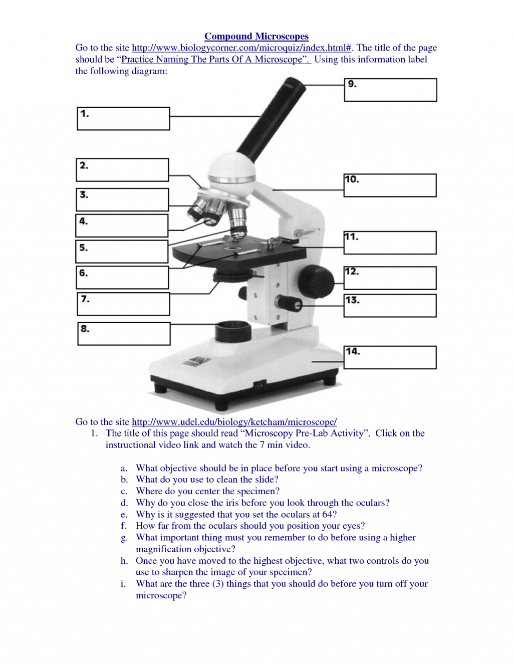
Using the Compound Microscope
07-843-6285 | 1/8 oz Tube | Terramycin Ophthalmic Ointment (Oxytetracycline HCl) An antibiotic possessing potent antimicrobial activity. It is one of the most versatile of the broad-spectrum antibiotics, and is effective in the treatment of infections due to gram-positive and gram-negative bacteria, bother aerobic and anaerobic, spirochetes, rickettsiae and certain of the larger viruses.

Cells and Microscopes
Compound Microscope: Definition, Diagram, Parts, Uses, Working ... - BYJUS A microscope with a high resolution and uses two sets of lenses providing a 2-dimensional image of the sample. The term compound refers to the usage of more than one lens in the microscope. Also, the compound microscope is one of the types of optical microscopes. The other type of optical microscope is a simple microscope.
Labeled Compound Microscope - ClipArt Best
10 Best Compound Microscopes (Summer 2022) - The Complete Guide Compound microscope is a type of optical microscope that is used for obtaining a high-resolution image. There are more than two lenses in a compound microscope. Learn about the working principle, parts and uses of a compound microscope along with a labeled diagram here.

How to Use a Compound Microscope: 6 Steps (with Pictures)
Electron microscope - Wikipedia An electron microscope is a microscope that uses a beam of accelerated electrons as a source of illumination. As the wavelength of an electron can be up to 100,000 times shorter than that of visible light photons, electron microscopes have a higher resolving power than light microscopes and can reveal the structure of smaller objects.. Electron microscopes use shaped magnetic …

What Is a Compound Microscope? | eHow
Microscope picture label Flashcards | Quizlet Start studying Microscope picture label. Learn vocabulary, terms, and more with flashcards, games, and other study tools.

Compound Light Microscope Labeled - Made By Creative Label
(PDF) Introduction to Microscopy - ResearchGate 08.11.2017 · • In compound microscope it will be i.e 10 X, f= 16 mm; 40 X, f= 4 mm; 100 X, f= 1.8 mm. • Image produced by objective lens falls on the eyepiece lens serve as objec t. …

OMAX MicroscopeNet: Phase Contrast Microscopy
Compound microscope - their parts and function - Microscopy4kids Compound microscopes have more than one lens to generate high magnification images of flat, thin specimens. 2. Eyepiece (10x) and Objective lenses (4x, 10x, 40x, 100x) are two major optical parts of a microscope. 3. Total magnification power is calculated by multiplying the magnification of the eyepiece and objective lens. 4.

What are the different types of microscopes used in biology > MISHKANET.COM
Amazing 27 Things Under The Microscope With Diagrams The tail is transparent and thus is difficult to detect under a low-power microscope. 23. Spirogyra under the microscope. Spirogyra is a green alga found mostly in freshwater in the form of green clumps. Spirogyra is unicellular, but because it clumps together, it can be seen in the pond even with our naked eyes.

Medical Technologists' Life in a Clinical Laboratory + Malaria Microscopy + Research ...
Looking at the Structure of Cells in the Microscope For deconvolution, we first obtain a series of (blurred) images, focusing the microscope in turn on a series of focal planes—in effect, a blurred three-dimensional image. The stack of images is then processed by computer to remove as much of the blur as possible. Essentially the computer program uses the microscope's point spread function to determine what the effect of the …

Before you buy a compound microscope | compoundmicroscopeblog
Tirth27/Skin-Cancer-Classification-using-Deep-Learning In the data pre-processing steps, all images are cropped into 768x786 and 512x512 resolution to reduce random noise on the edges of the image. The data cleaning and pre-processing step are performed on all the dataset obtained from the 2020, 2019 and 2018 competition. Also, the image labels are reconciled and combined into a single training CSV ...
template
What is a Compound Microscope? - New York Microscope Company A compound microscope is an instrument that is used to view magnified images of small specimens on a glass slide. It can achieve higher levels of magnification than stereo or other low power microscopes and reduce chromatic aberration. It achieves this through the use of two or more lenses in the objective and the eyepiece.

Compound Microscope Diagram | Microscope | Pinterest
Food Calorimetry: How to Measure Calories in Food We have the compound microscope you are looking for! Digital Microscopes. Digital microscopes are great for large classroom computer combined instruction. Students can take images, videos, and more. Stereomicroscopes. Stereomicroscopes show 3D images vs. flat images and are easier to focus and use. They are great for first tme student use. Physical & …
Post a Comment for "42 images of compound microscope with labels"