38 human eye with labels
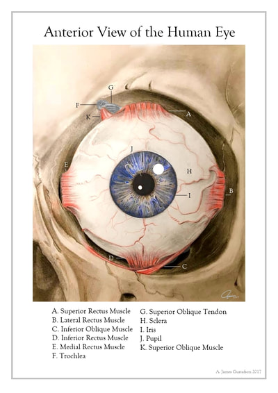
Eye Anatomy Labeling - Exploring Nature The style of citing shown here is from the MLA Style Citations (Modern Language Association). When citing a WEBSITE the general format is as follows. Author Last Name, First Name (s). "Title: Subtitle of Part of Web Page, if appropriate." Title: Subtitle: Section of Page if appropriate. Sponsoring/Publishing Agency, If Given. Parts of Human Eye and Their Functions | MD-Health.com The iris is the area of the eye that contains the pigment which gives the eye its color. This area surrounds the pupil, and uses the dilator pupillae muscles to widen or close the pupil. This allows the eye to take in more or less light depending on how bright it is around you. If it is too bright, the iris will shrink the pupil so that they ...
Draw a labeled sketch of the human eye - Class 8 Light - Teachoo The human eye is a large spherical ball which consists of Crystalline lens Aqueous Humour Pupil Iris Cornea Ciliary Muscles Retina Optic Nerve Vitreous Humour. Made by.
Human eye with labels
What Eye Problems Look Like - WebMD Warning Signs of Eye Trouble. 1 / 32. Blurry vision, spots, glare at night, flashing lights -- these are common eye complaints. Each could be a harmless annoyance or an early sign of disease. It ... The Iris: Anatomy, Function, and Treatment - Verywell Health The iris sits in the uveal tract, which is the eye's middle layer. The iris lies in front of the lens and behind the cornea. It is made up of the following parts: 13. Iris pigment epithelium contains melanin granules and chromatophores that make up the eye color. Dilator and sphincter muscles that expand and contract to control the amount of ... Understanding Different Eye Shapes: Which Do You Have? Hooded eyes: The lid appears smaller with this type of eye. This is because there is an extra layer of skin that is over the crease, which droops down. Protruding eyes: With this type of eye, the eyelids appear to project outward in the eye socket area. Upturned eyes: This type of eye has an almond shape.
Human eye with labels. All About the Eye Chart - American Academy of Ophthalmology The chart measures your visual acuity, or sharpness of vision. If you don't wear glasses or contacts, your eye doctor will use the results to find out whether you need them. If you wear corrective lenses, the results will show if your prescription needs to change. A standard Snellen vision testing chart from the 1950s. Eye Anatomy and Physiology a Complete Detail - Study Read It makes up the outermost part of eye anatomy. It is made of a dense, strong fibrous wall consisting of the sclera that is 5/6 th and the cornea that is anterior 1/6 th of the eyeball. The sclera is the outermost layer and it gives a definite shape to the eye. The gap between the sclera and the orbitals of the skull is filled with adipose tissue. Microscope Types (with labeled diagrams) and Functions A compound microscope: Is used to view samples that are not visible to the naked eye. Uses two types of lenses - Objective and ocular lenses. Has a higher level of magnification - Typically up to 2000x. Is used in hospitals and forensic labs by scientists, biologists and researchers to study micro organisms. Compound microscope labeled diagram. An Overview of the Eye's Iris - Verywell Health Overview of the Eye's Iris. The iris is the colored part of the eye that controls the amount of light that enters into the eye. It is the most visible part of the eye. The iris lies in front of the crystalline lens and separates the anterior chamber form the posterior chamber. The iris in part of the uveal tract which includes the ciliary body ...
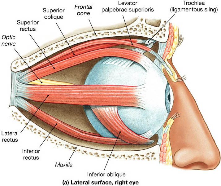
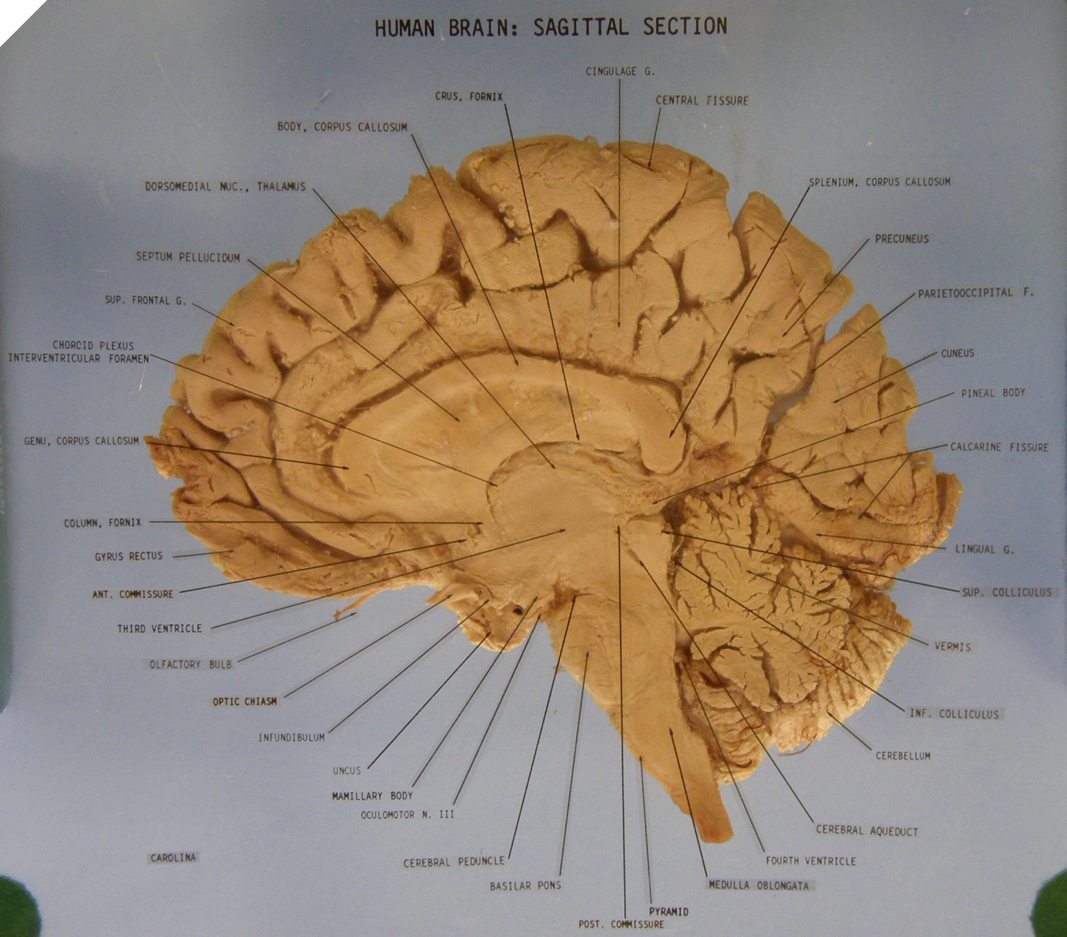
Brain: Atlas of human anatomy with MRI - e-Anatomy - IMAIOS Anatomy of the brain (MRI) - cross-sectional atlas of human anatomy. The module on the anatomy of the brain based on MRI with axial slices was redesigned, having received multiple requests from users for coronal and sagittal slices. The elaboration of this new module, its labeling of more than 524 structures on 379 MRI images in three different ... Visible Human Project: anatomy atlas of male cadaver This module presents the anatomy of the whole human body based on cross-sectional photographs of a male cadaver. 1300 anatomical structures have been labeled on 463 photographs of axial cross-sections. This atlas is based on the Visible Human Project ran by the U.S. National Library of Medicine (NLM) under the direction of Michael J. Ackerman. Eyeball: Structure and function | Kenhub The average human eye can see around 100 different shades of color and has a resolution that equals 576 gigapixels. These remarkable features of our eye are enabled by the complex structure of the eyeball. The eyeball consists of three layers; fibrous, vascular and nervous . Functionally, the most important layer is the retina, which receives ... Anatomy of the eye: Quizzes and diagrams | Kenhub How to learn the parts of the eye. Found within two cavities in the skull known as the orbits, the eyes are surrounded by several supporting structures including muscles, vessels, and nerves.There are 7 bones of the orbit, two groups of muscles (intrinsic ocular and extraocular), three layers to the eyeball… and that's just the beginning. There's a lot to learn, but stay calm!
Eye Diagram Quiz - ProProfs Try this amazing Eye Diagram Quiz quiz which has been attempted 4690 times by avid quiz takers. ... Label The Parts Of The Eye. People say that the eyes are the windows to a person's soul. ... How much did you get to understand about the human eye?... Questions: 8 | Attempts: 44609 | Last updated: Mar 22, 2022 . Sample Question. A is pointing ... Labeled imaging anatomy cases | Radiology Reference Article ... URL of Article. This article lists a series of labeled imaging anatomy cases by body region and modality. On this page: Article: Brain. Head and neck. Spine. Chest. Abdomen and pelvis. 40 Interesting Facts About Human Eyes A human eye can see 1 million colors. It also has an extraordinary ability to distinguish between different shades of a same color. For instance, human eyes can actually distinguish between 500 different gray shades. 19. The range of sight for human eyes is 200 degrees. 20. Only 1/6th of the entire eyeball is actually exposed to the outside ... Label-free imaging flow cytometry for analysis and sorting of ... For analysis of human photoreceptor cells, human retinal organoids (HRO) derived from the Crx-mCherry iPSC line (kindly provided by O. Goureau, Paris) and generated as described in Ref. 53 were ...
Human Anatomy
Neuroanatomy, Visual Pathway - StatPearls - NCBI Bookshelf Visual stimuli from our surroundings are processed by an intricate system of interconnecting neurons, which begins with the optic nerve in the eye up to the visual processing center in our forebrain called the visual cortex. All the information travels in the form of nerve impulses that are triggered by photosensitive chemical reactions occurring in the retina. Several separate and parallel ...
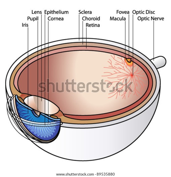
Human Eye Cross Section Labels Labels Stock Vector (Royalty Free) 89535880
Understanding Aqueous Humor and Vitreous Humor ... - NVISION Eye Centers The aqueous humor is a clear fluid located at the front part of the eye. Because the eye doesn't contain blood vessels, the aqueous humor is responsible for providing nutrients to the eye. The aqueous humor also drains out any excess material and waste from the eye. The vitreous fluid, or vitreous humor, is a colorless, transparent, gel-like ...
Human Eye Anatomy Images, Stock Photos & Vectors | Shutterstock
Understanding the Structure of the Eye - Lesson - TeachEngineering Students learn about the anatomical structure of the human eye and how humans see light, as well as some causes of color blindness. They conduct experiments as an example of research to gather information. During their investigations, they test other students' vision, gathering data and measurements about when objects appear blurry. These topics help students prepare to design solutions to an ...
picture front of the eye without labels clipart 20 free Cliparts | Download images on Clipground ...
Set of Human Sensory Organs: Anatomy of Five Sensory Organs, Eyes, Ear ... Five sense organs are equipped in the human body. Those organs provide us with first-hand information about our external or internal world. The names of those organs are stated below. Eye: perceives vision. Ear: perceives sound. Skin: perceives touch, temperature, roughness. Nose: perceives smell.
Chapter Two
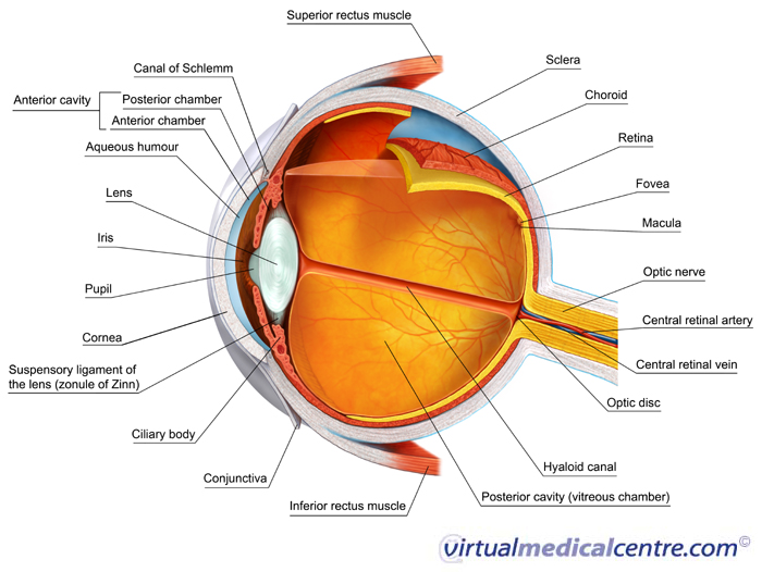
Anatomy of the Eye | BrightFocus Foundation Optic nerve: The bundle of nerve fibers at the back of the eye that carry visual messages from the retina to the brain. Photoreceptors: The light sensing nerve cells (rods and cones) located in the retina. Pupil: The adjustable opening at the center of the iris through which light enters the eye. Retina: The light sensitive layer of tissue that ...
(49).jpg)
How Well You Know The Human Eye? Trivia Quiz - ProProfs Quiz
Anatomy, Head and Neck, Face - StatPearls - NCBI Bookshelf The most anterior region of the head is the face. The human face is a unique aspect of each individual. The face contains many structures that contribute to the display of emotions, feeding, seeing, smelling, and communicating. One of the most distinguishing qualities of the face is that it is used for personal identity from person to person. Identity is essential since the face is usually the ...

Human Anatomy Lab: The Urinary and Reproductive Systems
External and internal eye anatomy - MedlinePlus Overview. The cornea allows light to enter the eye. As light passes through the eye the iris changes shape by expanding and letting more light through or constricting and letting less light through to change pupil size. The lens then changes shape to allow the accurate focusing of light on the retina. Light excites photoreceptors that ...

Human Anatomy Lab: Heart Models
Invisible machine-readable labels that identify and track objects Invisible machine-readable labels that identify and track objects ... new smartphone model with a camera that utilizes the infrared (IR) range of the electromagnetic spectrum that the naked eye can't perceive. ... Objects Using Low-Cost Infrared-Based 3D Printing and Imaging Tools," is being presented at the ACM CHI Conference on Human ...

Picture Of Human Eye With Labels - Human Anatomy
Quiz: Label The Parts Of The Eye - ProProfs People say that the eyes are the windows to a person's soul. In the class today, we covered parts of the eye, and what changes in them should be alarming to a patient. How much did you get to understand about the human eye? Take up this quiz and find out! Questions and Answers. 1.
The Anatomy of Human Eye with Diagram | EdrawMax Online
What color catches the human eye most? — COLORBUX The color that catches the human eye the most is either red or orange. Yellow is also a valid candidate, in some cases. Colors that are warm, bold, and bright are more eye-catching than others. Colors like red, orange, and yellow catch the human eye the most. This is why road signs and safety vests are made in these colors.
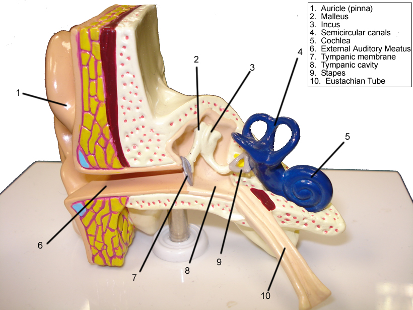
Eye and Ear Models
Glaucoma and the Importance of the Eye's Drainage System Glaucoma is a group of eye diseases that can damage the optic nerve and result in vision loss and blindness. In all forms of glaucoma, the status of the eye's drainage system, the trabecular meshwork, is important. Learn how the eye's drainage system relates to the development and treatment of glaucoma.

Human Anatomy Lab: Muscles of the Arm
Understanding Different Eye Shapes: Which Do You Have? Hooded eyes: The lid appears smaller with this type of eye. This is because there is an extra layer of skin that is over the crease, which droops down. Protruding eyes: With this type of eye, the eyelids appear to project outward in the eye socket area. Upturned eyes: This type of eye has an almond shape.

WMU Psychology Department: Lisa Baker
The Iris: Anatomy, Function, and Treatment - Verywell Health The iris sits in the uveal tract, which is the eye's middle layer. The iris lies in front of the lens and behind the cornea. It is made up of the following parts: 13. Iris pigment epithelium contains melanin granules and chromatophores that make up the eye color. Dilator and sphincter muscles that expand and contract to control the amount of ...

Amazing close-up photos of animal eyes (9 pics) | Amazing Creatures
What Eye Problems Look Like - WebMD Warning Signs of Eye Trouble. 1 / 32. Blurry vision, spots, glare at night, flashing lights -- these are common eye complaints. Each could be a harmless annoyance or an early sign of disease. It ...
Information on how to have healthy eyes | myVMC

How Your Eyes Work - Vision Center - Everyday Health
Post a Comment for "38 human eye with labels"