39 ear anatomy without labels
Parts of the Ear Labelled Diagram Activity - Twinkl The first worksheet presents an ear with annotations showing the first letters of its key features. For example, a label marked 'P' links to the Pinna (outer ear). The second page shows an ear diagram without labels. The final page shows the labels linking to the beginning letters of each feature, but without the words list. Label Anatomy Teaching Resources | Teachers Pay Teachers Anatomy Lab Cabinet Labels Room Decor - Script font. by. Teaching is a Lifestyle with Seeds of Study. $1.00. PDF. Human anatomy cabinet labels for anatomy models. Script font.Print on cardstock, cut out and stick to your cabinets. Each label is 4cmx18cmThe labels come with (and without) the word "system."
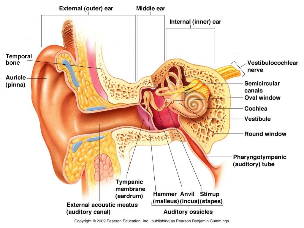
Picture of the Ear: Ear Conditions and Treatments - WebMD The ear has external, middle, and inner portions. The outer ear is called the pinna and is made of ridged cartilage covered by skin. Sound funnels through the pinna into the external auditory...

Ear anatomy without labels
Middle Ear Anatomy Stock Photos, Pictures & Royalty-Free ... Browse 422 middle ear anatomy stock photos and images available, or start a new search to explore more stock photos and images. Newest results. Human ear anatomy. Ears inner structure, Medical Education Chart of Biology,Human ear in medical concept, anatomical structure,3D rendering Human ear anatomy. Anatomy of the Ear | Inner Ear | Middle Ear | Outer Ear Anatomy of the Ear. The ear is made up of three parts: the outer, middle, and inner ear. All three parts of the ear are important for detecting sound by working together to move sound from the outer part through the middle and into the inner part of the ear. Ears also help to maintain balance. Ear Anatomy Without Labels Digital Art Stock Illustration ... Ear Anatomy Without Labels Digital Art Stock Illustration 530108302 Edit Download for free See more Popularity score High Usage score High usage Superstar Shutterstock customers love this asset! Item ID: 530108302 Ear Anatomy without Labels, Digital Art Formats 8976 × 6201 pixels • 29.9 × 20.7 in • DPI 300 • JPG
Ear anatomy without labels. Human Body Parts Images Without Labels - Free Vector ... Human ear diagram with labels and label of anatomy labeling the ear purposegames nose diagram with label diagrams all labels human ear the ear diagram without labels anatomy human charts. Illustration Of Body Parts Labels It is certainly the most widely studied structure the world over. Human body parts images without labels. Download body ... Blank ear diagrams and quizzes: The fastest way to learn ... Ear diagrams (labeled and unlabeled) Accelerate your learning with interactive quizzes Sources + Show all Ear anatomy overview Although it's not obvious to look at, the ear is anatomically divided into three portions: External (outer) ear Middle ear Inner ear As you can imagine, there's a lot of associated anatomy to learn for each portion! Human Eye Diagram Without Labels - solved label this ... Human Eye Diagram Without Labels - 13 images - picture front of the eye without labels clipart clipground, diagram of the eye clipart etc, eye diagram images stock photos vectors shutterstock, eye anatomy images stock photos vectors shutterstock, Well-Labelled Diagram Of Ear With Explanation - BYJUS Eustachian Tube is a tube that connects the middle ear to the back of the nose. It helps to maintain equal pressure in the middle ear which facilitates the proper transmission of sound waves. The Inner ear consists of: Cochlea that comprises the nerves of hearing. Semicircular canals that contain the receptors that help in maintaining balance.
File:Ear-anatomy-text-small-en.svg - Wikipedia File:Ear-anatomy-text-small-en.svg. Size of this PNG preview of this SVG file: 790 × 599 pixels. Other resolutions: 317 × 240 pixels | 633 × 480 pixels | 1,013 × 768 pixels | 1,280 × 971 pixels | 2,560 × 1,942 pixels | 1,450 × 1,100 pixels. This is a file from the Wikimedia Commons. Information from its description page there is shown below. Outer Ear Anatomy Colorful Pattern Without Center Label ... Shop "outer ear anatomy colorful pattern without center label" search results for the very best in custom shoes, sneakers, apparel, and accessories by independent artists. Label Parts of the Human Ear - University of Dayton Label Parts of the Human Ear. Select One Auditory Canal Cochlea Cochlear Nerve Eustachian Tube Incus Malleus Oval Window Pinna Round Window Semicircular Canals Stapes Tympanic Membrane Vestibular Nerve. Select One Auditory Canal Cochlea Cochlear Nerve Eustachian Tube Incus Malleus Oval Window Pinna Round Window Semicircular Canals Stapes ... Human Ear Diagram - Bodytomy Auditory Ossicles: The three small bones in the middle ear, called malleus, stapes, and incus, are connected. These bones together are called the auditory ossicles, and their purpose is to let the sound that strikes the eardrum, further into the inner ear.
The Ear: Anatomy, Function, and Treatment - Verywell Health Essential organs of human hearing and balance, the ears are located on either side of the head, at the level of the nose. Separated into an inner, middle, and outer ear, each ear is an intricate and complicated mixture of bones, nerves, and muscles. Ear Diagram and Labeling Worksheet / Worksheet The first worksheet presents an ear with annotations showing the first letters of its key features. For example, a label marked 'P' links to the Pinna (outer ear). The second page shows an ear diagram without labels. The final page shows the labels linking to the beginning letters of each feature, but without the words list. Image result for ear structure without label | Ear diagram ... Feb 12, 2018 - Image result for ear structure without label. Feb 12, 2018 - Image result for ear structure without label. Pinterest. Today. Explore. ... Label Ear Anatomy Diagram Printout. G. Kim Gregory. Science. Human Brain Anatomy. Anatomy And Physiology. Animal Coloring Pages. Coloring Book Pages. Ear Anatomy: Understanding the Outer, Middle, and Inner ... The external auditory meatus, or ear canal, is a narrow canal that leads from the concha to the tympanic membrane, or eardrum. Sound waves are delivered through this canal. This canal is prone to ear infections. Tragus This is the small, rigid part of the ears along the front of the ear, adjacent to the face.
Ear Anatomy Name With Pictures: | All In One About Medical
Human Ear Anatomy - Parts of Ear Structure, Diagram and ... The external (outer) ear consists of the auricle, external auditory canal, and eardrum (Figure 1 and 2). The auricle or pinna is a flap of elastic cartilage shaped like the flared end of a trumpet and covered by skin. The rim of the auricle is the helix; the inferior portion is the lobule. Ligaments and muscles attach the auricle to the head.
Hearing Loss, Tinnitus & Balance Therapy | Pacific Eye & Ear Center
Outer Ear: Anatomy, Location, and Function - Verywell Health Helix: The outermost curvature of the ear, extending from where the ear joins the head at the top to where it meets the lobule. The helix begins the funneling of sound waves into the ear; Fossa, superior crus, inferior crus, and antihelix: These sections make up the middle ridges and depressions of the outer ear. The superior crus is the first ridge that emerges moving in from the helix.
Functioning of the Human Ear - Science Wiz
Bones Of The Human Body Without Labels at Anatomy Bones Of The Human Body Without Labels. They range in size from the tiniest found in the middle ear, to the largest that forms our thigh.the human body has an amazing array of different bones, many of which you can find on yourself or on a skeleton.knowledge of the skeletal structure of the human body is essential to know before.

The Heart - Science Quiz
Ear pain - Wikipedia Ear pain, also known as earache or otalgia, is pain in the ear. Primary ear pain is pain that originates from the ear. Secondary ear pain is a type of referred pain, meaning that the source of the pain differs from the location where the pain is felt. . Most causes of ear pain are non-life-threatening. Primary ear pain is more common than secondary ear pain, and it is often due to infection or ...
Anatomy of Ear – Anatomy
Anatomy of the Ear | Geeky Medics The tympanic membrane, or eardrum, marks the border between the external and middle ear. It is formed of a middle layer of connective tissue with a layer of skin on its lateral surface (facing the external acoustic meatus) and mucous membrane on its medial surface (facing the middle ear).

Unlabeled Eye Diagram — UNTPIKAPPS
Ear Anatomy Diagram - EnchantedLearning.com Ear Anatomy: Ear Anatomy: Label Me! Printout: Sound is collected by the pinna (the visible part of the ear) and directed through the outer ear canal. The sound makes the eardrum vibrate, which in turn causes a series of three tiny bones (the hammer, the anvil, and the stirrup) in the middle ear to vibrate. The vibration is transferred to the ...

Trigger Points Set Exam-Room Anatomy Posters – ClinicalPosters
Skeletal System Diagram Without Labels - Anatomy Organ Apr 5, 2018 - Skeletal System Diagram Without Labels - See more about Skeletal System Diagram Without Labels, skeletal system diagram no labels, skeletal system diagram without labels, skeletal system picture without label

Ear Anatomy Flashcards | Quizlet
Ear Anatomy - Outer Ear | McGovern Medical School The medical term for the outer ear is the auricle or pinna. The outer ear is made up of cartilage and skin. There are three different parts to the outer ear; the tragus, helix and the lobule. EAR CANAL The ear canal starts at the outer ear and ends at the ear drum. The canal is approximately an inch in length.

The Eye - Science Quiz
Ear Anatomy Without Labels Digital Art Stock Illustration ... Ear Anatomy Without Labels Digital Art Stock Illustration 530108302 Edit Download for free See more Popularity score High Usage score High usage Superstar Shutterstock customers love this asset! Item ID: 530108302 Ear Anatomy without Labels, Digital Art Formats 8976 × 6201 pixels • 29.9 × 20.7 in • DPI 300 • JPG

Diagram Of Human Ear With Labelling - Human Anatomy
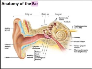
Anatomy of the Ear | Inner Ear | Middle Ear | Outer Ear Anatomy of the Ear. The ear is made up of three parts: the outer, middle, and inner ear. All three parts of the ear are important for detecting sound by working together to move sound from the outer part through the middle and into the inner part of the ear. Ears also help to maintain balance.
Anatomy of the ear
Middle Ear Anatomy Stock Photos, Pictures & Royalty-Free ... Browse 422 middle ear anatomy stock photos and images available, or start a new search to explore more stock photos and images. Newest results. Human ear anatomy. Ears inner structure, Medical Education Chart of Biology,Human ear in medical concept, anatomical structure,3D rendering Human ear anatomy.

Diagram Of Human Ear With Labelling - Human Anatomy
picture front of the eye without labels clipart 20 free Cliparts | Download images on Clipground ...
FREE Ear Anatomy Printable - Homeschool Giveaways
Post a Comment for "39 ear anatomy without labels"